Low Back Pain: Red Flags, Imaging Rules, and Exercise Therapy
 Sep, 4 2026
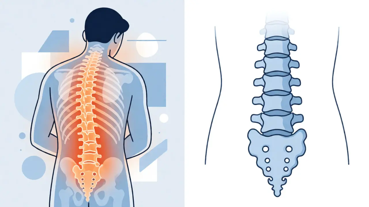
Here is a number that might make you pause: about 80% of adults will deal with low back pain at some point in their lives. It’s the second most common reason people visit a doctor in the United States, right behind colds and flu. But here is the twist that surprises most patients: for roughly 97% of those cases, there is no serious disease hiding in your spine. It’s mechanical. It’s annoying. It hurts. But it isn’t dangerous.
The real challenge isn’t just managing the ache; it’s knowing when to worry. Should you rush to an MRI? Do you need surgery? Or can you fix this by moving differently? This guide cuts through the noise using current clinical guidelines from the American College of Physicians and the American Academy of Family Physicians. We’ll break down exactly which "red flags" actually matter, why early imaging often does more harm than good, and how specific exercises are proven to work better than pills for long-term relief.
The Reality of Low Back Pain Statistics
If you’re reading this because your back currently feels like it’s been hit by a truck, you aren’t alone. Data from the Global Burden of Disease Study shows that low back pain is the leading cause of disability worldwide. Yet, despite its prevalence, our understanding of it has shifted dramatically over the last two decades.
In the past, doctors often treated every bad back as a structural failure-a slipped disc or a broken vertebra that needed fixing. Today, we know that structure doesn’t always equal symptoms. You could have a herniated disc on an MRI and feel fine, while someone else with a "clean" scan is in agony. This disconnect is why modern medicine focuses less on finding a single anatomical culprit and more on functional recovery.
Most episodes are acute, meaning they last less than four weeks. About one-third of people still have some discomfort after three months, turning it into chronic pain. The goal of treatment isn’t necessarily to eliminate all sensation immediately-that’s unrealistic-but to restore function and prevent recurrence. Understanding this context helps lower anxiety, which itself plays a huge role in how much pain you actually feel.
Red Flags: When to Worry (And When Not To)
Doctors use a checklist called "red flags" to screen for the rare 1-2% of cases where low back pain signals something serious, like cancer, infection, fracture, or nerve compression. But not all red flags are created equal. Some are strong indicators; others are weak clues that lead to unnecessary panic and testing.
A major review published in the European Spine Journal identified 46 different red flags across various guidelines, but only two appear consistently in more than 75% of them: a history of cancer and major trauma. Everything else is murkier. For instance, age over 50 was once considered a standalone warning sign. However, recent evidence suggests age alone has a positive likelihood ratio of just 1.2 for serious pathology-barely better than a coin flip. So, if you’re 55 with a sore back but no other symptoms, you likely don’t need an emergency workup.
Let’s look at the specific categories and what actually matters:
- Cauda Equina Syndrome: This is a true emergency. It involves compression of the nerve roots at the bottom of the spinal cord. Key signs include bladder or bowel incontinence, urinary retention (inability to pee), saddle anesthesia (numbness in the groin/buttocks area), and loss of anal sphincter tone. If you have these, go to the ER immediately. Studies show surgical decompression within 48 hours yields significantly better outcomes.
- Fracture: Major trauma (like a car accident or fall from height) is a clear flag. Also, consider steroid use or osteoporosis risk factors. Minor strains from lifting groceries rarely cause fractures unless bones are already compromised.
- Malignancy (Cancer): A known history of cancer is the strongest predictor. Unintentional weight loss and night pain (pain that wakes you up and doesn’t change with position) are also relevant, though less specific.
- Infection: Look for fever, chills, IV drug use, or recent invasive procedures. Vertebral tenderness when tapping the spine can be a clue, but it’s not definitive without blood tests like ESR or CRP.
Notice what isn’t on this list: tingling in the legs alone, muscle spasms, or stiffness in the morning. These are typical mechanical symptoms. Misinterpreting them as red flags leads to what experts call a "diagnostic cascade"-one test leads to another, costing billions annually without improving health outcomes.
Imaging: Why Your MRI Might Be Overrated
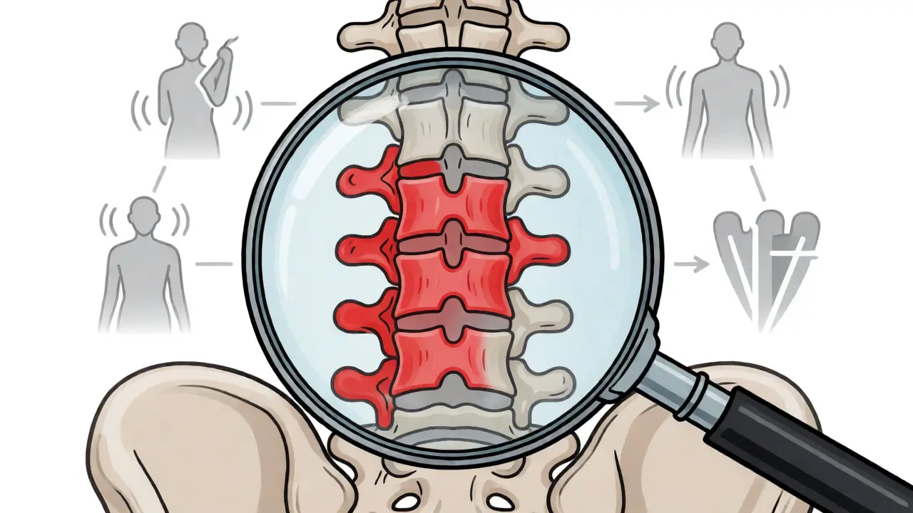
We live in an era where we expect answers from technology. If your knee hurts, you want an X-ray. If your back hurts, you want an MRI. But for non-specific low back pain, guidelines strongly advise against routine imaging in the first six weeks unless red flags are present.
Why? Because findings on imaging often don’t correlate with pain levels. An MRI might show disc degeneration or bulging discs in healthy, pain-free individuals. Seeing these abnormalities can create a "nocebo" effect-you start believing your back is broken, which increases fear and avoidance behaviors, ultimately worsening pain.
The American College of Radiology Appropriateness Criteria clarifies this well:
| Scenario | Recommended Action | Rationale |
|---|---|---|
| Acute pain (<4 weeks), no red flags | No imaging | High rate of spontaneous resolution; imaging adds cost/radiation without changing management. |
| Suspected Cauda Equina Syndrome | MRI Lumbar Spine | Urgent need to visualize nerve root compression for potential surgery. |
| Suspected Fracture/Trauma | X-ray or CT | Bone detail is best seen on CT/X-ray; faster and cheaper than MRI for bone issues. |
| Suspected Infection/Cancer | MRI with contrast or Bone Scan | Soft tissue involvement and marrow changes require high-resolution soft-tissue imaging. |
For subacute or chronic pain without red flags, even X-rays have limited utility. They show bones, not discs or nerves. Unless you suspect arthritis or spondylolisthesis (slipped vertebra), an X-ray rarely changes the treatment plan. Save your money and radiation exposure. Focus instead on clinical assessment: how does movement affect your pain? What makes it better or worse?
Exercise Therapy: The Gold Standard Treatment
If imaging is often unnecessary, what is necessary? Movement. Specifically, structured exercise therapy. This isn’t about running marathons or heavy deadlifts on day one. It’s about targeted rehabilitation that restores control, strength, and endurance to your core and hip muscles.
A massive Cochrane Review analyzing nearly 20,000 participants confirmed that exercise therapy produces statistically significant reductions in pain and improvements in function compared to no treatment. The effect sizes are modest but clinically meaningful, especially for long-term prevention. More importantly, active treatment beats passive treatments like massage, heat packs, or ultrasound for lasting results.
Not all exercises are equal. Research highlights three approaches with the strongest evidence:
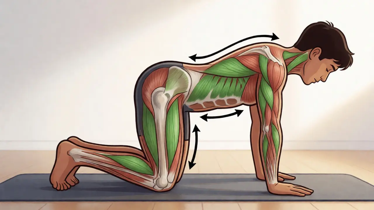
- Motor Control Exercises: These focus on retraining deep stabilizing muscles, particularly the transverse abdominis and multifidus. Think pelvic tilts, bird-dogs, and bridges. The goal is teaching your nervous system to stabilize your spine during daily tasks.
- Graded Activity: This involves breaking activities into small, manageable steps. Instead of trying to lift a box all at once, you practice the motion repeatedly with light weights, gradually increasing load. It builds confidence and tolerance.
- Combined Programs: Mixing aerobic exercise (walking, swimming, cycling) with strengthening yields the best results. Aerobic activity improves blood flow and reduces systemic inflammation, while strength training supports the skeletal structure.
The American Physical Therapy Association recommends starting with 2-3 supervised sessions per week for 8-12 weeks. Adherence is key. If you quit after two weeks, you won’t see benefits. Start low and slow. Aim for 40-60% of your maximum effort initially, progressing to 70-85% as pain allows. Consistency trumps intensity every time.
Practical Pitfalls and How to Avoid Them
Even with good intentions, patients often fall into traps that delay recovery. Here are the most common ones based on clinical experience and patient surveys.
Pitfall 1: Bed Rest. Old advice said rest until it heals. New advice says keep moving. Prolonged bed rest weakens muscles, stiffens joints, and increases depression scores. Aim to stay active within pain limits. Walking is usually safe and beneficial.
Pitfall 2: Fear-Avoidance Behavior. If you believe any movement will damage your spine, you’ll move less. Less movement leads to deconditioning, which leads to more pain. Break this cycle by recognizing that hurt does not equal harm. Discomfort during gentle exercise is normal and often safe.
Pitfall 3: Relying Solely on Passive Treatments. Chiropractic adjustments or physical therapy modalities (TENS units, ice) can provide short-term relief. That’s okay! But they should serve as a bridge to active rehab, not the destination. Use pain relief to enable exercise, not replace it.
Pitfall 4: Ignoring Psychosocial Factors. Stress, anxiety, poor sleep, and job dissatisfaction amplify pain perception. Addressing these through mindfulness, cognitive behavioral techniques, or simply improving sleep hygiene can reduce back pain intensity as effectively as medication.
When to Seek Immediate Help
While most back pain resolves with self-care and exercise, certain scenarios demand urgent attention. Don’t wait if you experience:
- Loss of bladder or bowel control.
- Numbness in the inner thighs or genital area.
- Progressive weakness in both legs (e.g., foot drop).
- Fever combined with severe back pain.
- Severe pain following significant trauma.
For everything else, give it time. Most acute episodes improve within 4-6 weeks. If pain persists beyond that, consult a primary care provider or physical therapist for a tailored plan. Remember, you are the CEO of your body. Ask questions. Challenge unnecessary tests. And prioritize movement over medication whenever possible.
Do I need an MRI for my lower back pain?
Generally, no. Guidelines recommend against imaging for acute low back pain (under 4-6 weeks) unless you have "red flags" such as progressive neurological deficits, suspected cancer, infection, or cauda equina syndrome. Early MRIs often reveal incidental findings that don’t explain your pain and may lead to unnecessary treatments.
What are the most reliable red flags for serious back conditions?
The strongest red flags are a history of cancer, major trauma (like a fall from height or car accident), unexplained weight loss, fever, and signs of cauda equina syndrome (bladder/bowel dysfunction, saddle anesthesia). Age alone is a weak predictor and shouldn’t trigger immediate imaging by itself.
How long should I do exercise therapy for back pain?
Clinical guidelines suggest a minimum of 8-12 weeks of structured exercise therapy for significant improvement. This typically involves 2-3 supervised sessions per week initially, transitioning to a home program. Consistency is crucial; stopping early often leads to recurrence.
Is bed rest good for acute low back pain?
No. Current evidence advises against prolonged bed rest. Staying active within pain limits speeds up recovery. Short periods of rest (1-2 days) may help with severe acute pain, but returning to normal activities as soon as possible prevents muscle weakening and stiffness.
Can stress cause lower back pain?
Yes. Psychological factors like stress, anxiety, and fear of movement can amplify pain perception and contribute to chronicity. Managing stress through mindfulness, adequate sleep, and cognitive-behavioral strategies is an important part of comprehensive back pain care.
Jeremy Westcott
September 5, 2026 AT 18:38They don't want you to know this, but the whole 'mechanical pain' narrative is a smokescreen to keep us from questioning why our spines are literally crumbling under the weight of modern life. It's not just about moving; it's about the toxic environment we're forced to inhabit. They push pills and MRIs because they profit off your fear, not your healing. Wake up.
Amanda SF
September 6, 2026 AT 17:03I found the article rather pedestrian in its approach.
While the statistics cited are accurate, the tone lacks the gravitas one expects when discussing a condition that afflicts nearly every living soul at some point. The advice on imaging is sound, yes, but the dismissal of passive treatments feels overly simplistic for such a complex physiological phenomenon. One must consider the psychological toll before rushing into 'movement therapy.' It is all rather tedious, frankly.
Neil Martin
September 7, 2026 AT 17:41The distinction between nociceptive input and actual tissue damage is critical here, yet most patients fail to grasp the biopsychosocial model entirely. We see a massive over-reliance on anatomical determinism which leads to unnecessary iatrogenic harm through diagnostic cascades. If clinicians aren't properly screening for psychosocial yellow flags alongside red flags, they are practicing substandard care. It’s frustrating to watch people chase structural fixes for functional problems.
Kim Pender
September 9, 2026 AT 15:08Finally someone says it. Stop getting MRIs for everything. I waited six weeks and my back healed itself. No surgery needed.
Neil Sahli
September 9, 2026 AT 21:17Love the energy here! But let's be real, telling people to 'just move' when they're in agony is like telling a drowning person to swim better. You gotta ease them in. Start with breathing, then walking, THEN the core work. Otherwise, they quit day three and blame the exercise, not their expectation management. Keep pushing that consistency message though, it's gold!
Pramay Dattani
September 11, 2026 AT 11:24In India we dont have this luxury of waiting 6 weeks. Our doctors rush to MRI immediatly becuase they think they know best. But actually its waste of money. Also you forgot to mention yoga? Its better than western exercises for spine health. Dont ignore ancient wisdom.
Falgun R Patel
September 12, 2026 AT 08:45This resonates deeply with me. We often look for external solutions-scans, surgeries, medications-when the path to recovery lies within our own relationship with movement and perception. The idea that 'hurt does not equal harm' is a philosophical shift as much as a medical one. It requires patience and self-compassion. To those suffering, remember that your body is trying to protect you, not punish you. Listen gently.
Jim Bisesi
September 14, 2026 AT 02:05meh. good info but too long. nobody reads this much.