Keratoconus Management: How Rigid Lenses Restore Vision in Progressive Corneal Thinning
 Sep, 2 2026
Imagine trying to read a newspaper through the bottom of a wine glass. The text is distorted, stretched, and impossible to focus on. That’s what living with Keratoconus feels like for many patients. It isn’t just blurry vision; it’s a structural collapse of the eye’s window. The cornea, normally a perfect dome, bulges outward into a cone shape due to progressive thinning. This condition affects roughly 1 in 2,000 people worldwide, often striking during teenage years or early adulthood. While glasses can help in the earliest stages, they fail once the irregularity sets in. This is where rigid lenses step in as the gold standard for non-surgical visual rehabilitation.
| Aspect | Detail |
|---|---|
| Nature of Disease | Progressive thinning and bulging of the cornea (cone shape). |
| Primary Symptom | Distorted, wavy vision that worsens over time; light sensitivity. |
| Standard Solution | Rigid Gas Permeable (RGP) or Scleral lenses create a smooth optical surface. |
| Disease Halt | Corneal Cross-Linking (CXL) stops progression but doesn't fix existing distortion. |
| Adaptation Time | Typically 2-4 weeks for full-time wear comfort. |
Why Your Cornea Is Changing Shape
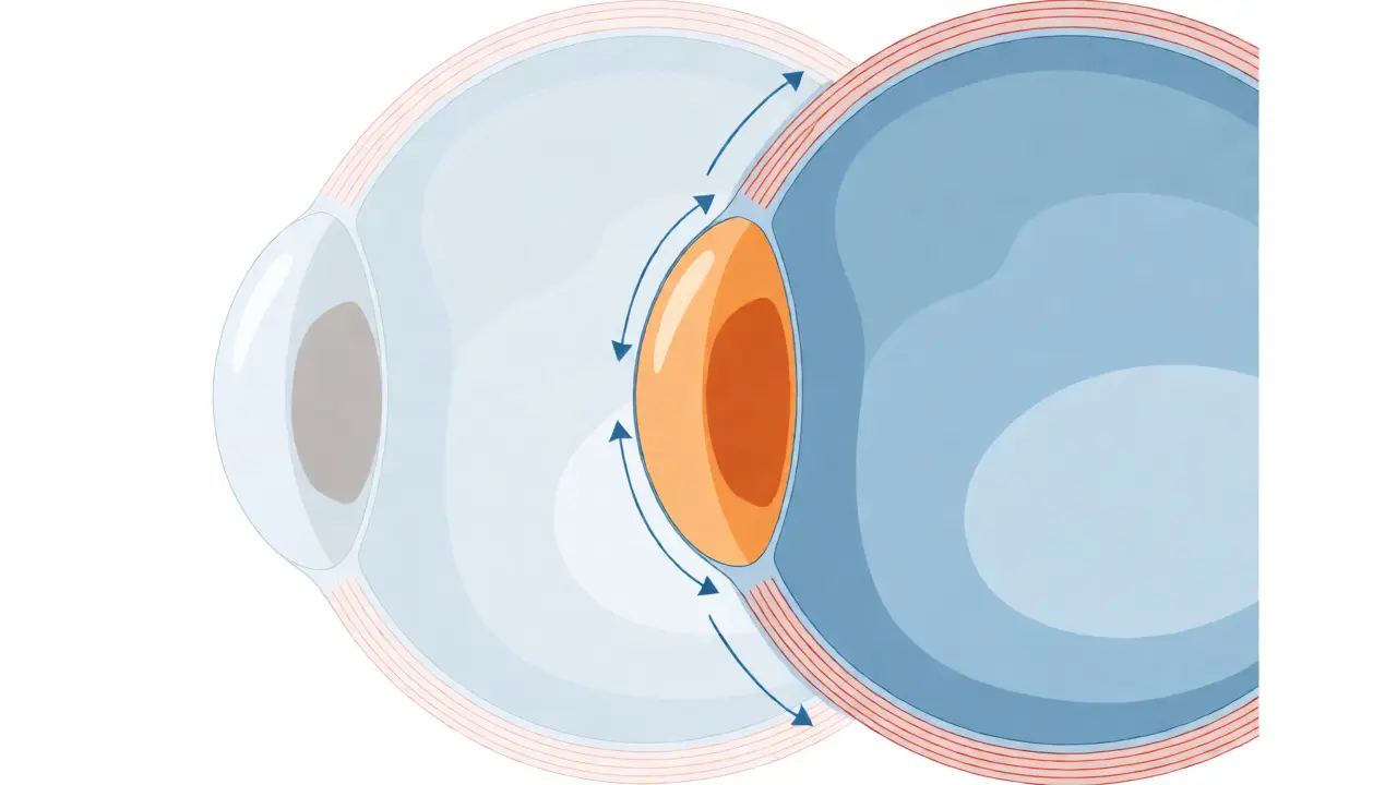
To understand why rigid lenses are necessary, you have to look at what’s happening inside your eye. Keratoconus is defined as a progressive central or inferocentral corneal thinning and irregularity. Think of the cornea as a balloon. If one spot becomes weak, air pressure pushes it out, creating a bulge. In keratoconus, this weakness stems from an imbalance in enzymes. There is a marked increase in degradative enzymes and a decrease in their inhibitors, which breaks down the collagen structure holding the cornea together. According to clinical guidelines from Wills Eye Hospital, this process almost always affects both eyes, though rarely equally-one eye is usually worse than the other.
The damage isn't random. It follows a gradient pattern. The microscopic keratocytes (cells in the cornea) penetrate specific layers, causing structural failure. Topography scans reveal a distinct "steepening" pattern, particularly in the inferior-central region. This creates the classic cone shape. Without intervention, this thinning continues until the fourth decade of life, when the disease typically stabilizes. But stabilization doesn't mean restoration. Once the cone forms, your vision remains distorted unless corrected externally.
The Physics of Rigid Lenses
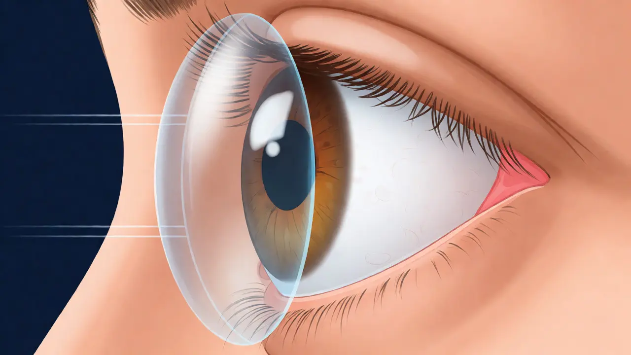
Soft contact lenses mold to the shape of your eye. If your eye is bumpy, the soft lens gets bumpy too. They simply cannot correct the irregular astigmatism caused by keratoconus. Rigid Gas Permeable (RGP) lenses work differently. They are made of stiff materials that maintain their own spherical shape regardless of the cornea underneath. When placed on the eye, tears fill the gap between the rigid lens and the irregular cornea. This tear layer acts as a new, perfectly smooth refractive surface. Light enters through the rigid lens and the fluid reservoir, bypassing the chaotic corneal surface entirely.
This mechanism provides clarity that glasses simply cannot match. Studies from Northwest Eye Clinic show visual acuity improvements from a median of 20/400 to 20/200 post-fitting in moderate cases. For advanced cases, the jump is even more dramatic. Patients report a significant reduction in ghosting and halos around lights, which are common complaints with spectacle correction in keratoconus. The key here is rigidity. The lens must be stiff enough to vault the cone without touching it, yet small enough to stay centered on the eye.
Choosing Between RGP and Scleral Lenses
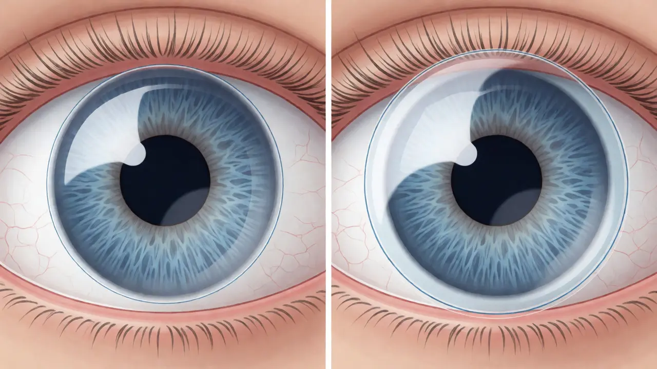
Not all rigid lenses are created equal. As the disease progresses, traditional RGPs might become uncomfortable or unstable. This is where Scleral Lenses come into play. These are larger, ranging from 15mm to 22mm in diameter. Instead of resting on the sensitive cornea, scleral lenses vault over the entire corneal surface and rest on the white part of the eye, the sclera. This design creates a large fluid reservoir that hydrates the cornea continuously.
For patients with extreme irregularity or dry eye issues, sclerals are often the only viable option. Dr. Gregory Kent of Boise LASIK Center notes that scleral lenses represent a viable option for those struggling with vision loss due to advanced keratoconus when other treatments fail. The success rate for scleral lenses in stage III-IV keratoconus is approximately 85%, compared to 65% for traditional RGPs. However, fitting them is complex. It requires specialized topography equipment and multiple visits-often 3 to 5 over 4 to 6 weeks-to get the parameters right. Manufacturers like BostonSight offer customized designs like PROSE lenses, which use digital manufacturing based on individual eye scans.
Stopping Progression vs. Correcting Vision
A common misconception is that wearing rigid lenses cures keratoconus. They don't. They correct vision. To stop the disease from getting worse, you need a different tool: Corneal Cross-Linking (CXL). FDA-approved in 2016, CXL uses riboflavin drops and UV light to strengthen the collagen bonds in the cornea. It has a 90-95% success rate in halting progression over five years. But here’s the catch: CXL locks the cornea in its current shape. If you wait until your vision is severely distorted to get CXL, you’ll still need rigid lenses afterward to see clearly.
Most specialists now recommend a combined approach. You halt the progression first with CXL, then rehabilitate your vision with rigid lenses. Ignoring this dual strategy leads to frustration. Patients often ask if they can skip CXL and just wear lenses. You can, but if the disease progresses, your lens prescription will change frequently, and eventually, the cornea may scar or thin so much that lenses no longer fit. At that point, you’re looking at surgery.
The Reality of Adaptation
Let’s be honest: putting a hard plastic disc on your eye feels weird at first. About 30% of patients experience initial discomfort. Common complaints include foreign body sensation (reported by 45% of new wearers), lens awareness (38%), and difficulty with insertion (32%). This is normal. Your brain needs time to ignore the physical presence of the lens while processing the sharp image it provides.
Successful adaptation follows a predictable curve. Start with 2-4 hours of daily wear. Increase by 1-2 hours each day. Most patients achieve comfortable full-time wear within 2-4 weeks. During this period, you’ll learn proper hygiene protocols. Daily cleaning and disinfection are non-negotiable. Skipping steps can lead to infections or giant papillary conjunctivitis, an allergic reaction to deposits on the lens. Long-term users report high satisfaction, citing improved confidence and reduced dependency on others for reading or driving. The trade-off is effort. You are trading convenience for clarity.
When Surgery Becomes Necessary
Rigid lenses are powerful, but they aren't magic. Approximately 10-20% of keratoconus patients eventually require a corneal transplant because they cannot tolerate lenses or have advanced scarring. There are two main types: Penetrating Keratoplasty (PK), which replaces the entire cornea, and Deep Anterior Lamellar Keratoplasty (DALK), which replaces only the front layers, preserving the healthy back endothelium. DALK generally has a lower rejection risk.
Even after a transplant, vision isn't instant. Rehabilitation can take over 12 months. Furthermore, graft rejection occurs in 5-10% of cases. Some patients also explore INTACS-implantable ring segments that flatten the cone. Approved in 1999, INTACS can reduce myopia and astigmatism, potentially allowing some patients to switch back to glasses or softer lenses. However, 35-40% of INTACS recipients still end up needing rigid lenses for optimal vision. The hierarchy of treatment usually flows from glasses → rigid lenses + CXL → INTACS/Sclerals → Transplant.
Troubleshooting Common Issues
If you’re starting rigid lens therapy, expect a few bumps in the road. Lens decentration (the lens sliding off-center) happens in about 15% of cases, often due to eyelid tension or improper base curve. Fogging, reported by 25% of users, is usually a sign of poor oxygen exchange or dirty lenses. Switching to higher oxygen permeability materials (Dk values >100) or preservative-free rewetting drops can solve this.
Another frequent issue is solution sensitivity. Some patients react to preservatives in standard contact lens solutions. Using hydrogen peroxide-based systems or preservative-free saline rinses often clears up redness and irritation. Remember, if pain accompanies redness, remove the lens immediately and call your doctor. Pain is not part of the adaptation process; it’s a warning sign of abrasion or infection.
Can I wear regular soft contacts for keratoconus?
In very mild, early-stage keratoconus, specialty soft toric lenses might provide acceptable vision. However, as the cone develops, soft lenses conform to the irregular cornea rather than masking it. They typically fail to correct the high amounts of irregular astigmatism, leading to blurred and distorted vision. Rigid lenses are superior because they create a new, smooth optical surface via the tear layer beneath them.
How long does it take to get used to rigid lenses?
Most patients adapt within 2 to 4 weeks. The process involves gradually increasing wear time, starting with 2-4 hours a day and adding 1-2 hours daily. Initial sensations of foreign body feeling or awareness are normal and usually subside as the brain learns to ignore the lens. Full-time comfort is achieved by 85% of patients who persist through this period.
Do rigid lenses stop keratoconus from getting worse?
No, rigid lenses do not stop the progression of the disease. They only correct the vision impairment caused by the irregular corneal shape. To halt progression, Corneal Cross-Linking (CXL) is required. Many patients undergo CXL to stabilize the cornea and then wear rigid lenses to manage the residual visual distortion.
What is the difference between RGP and scleral lenses?
RGP (Rigid Gas Permeable) lenses are smaller (9-10mm) and rest directly on the cornea. They are suitable for mild to moderate cases. Scleral lenses are larger (15-22mm) and vault over the cornea, resting on the sclera (white of the eye). They hold a reservoir of fluid against the cornea, providing better hydration and stability for advanced cases or patients with dry eyes. Sclerals are generally more comfortable but require a more complex fitting process.
Is corneal transplant inevitable for keratoconus patients?
No. Only about 10-20% of patients ultimately require a corneal transplant. Advances in cross-linking and specialty contact lenses (especially sclerals) have significantly reduced the need for surgery. Most patients manage their condition successfully with lenses and monitoring for decades.